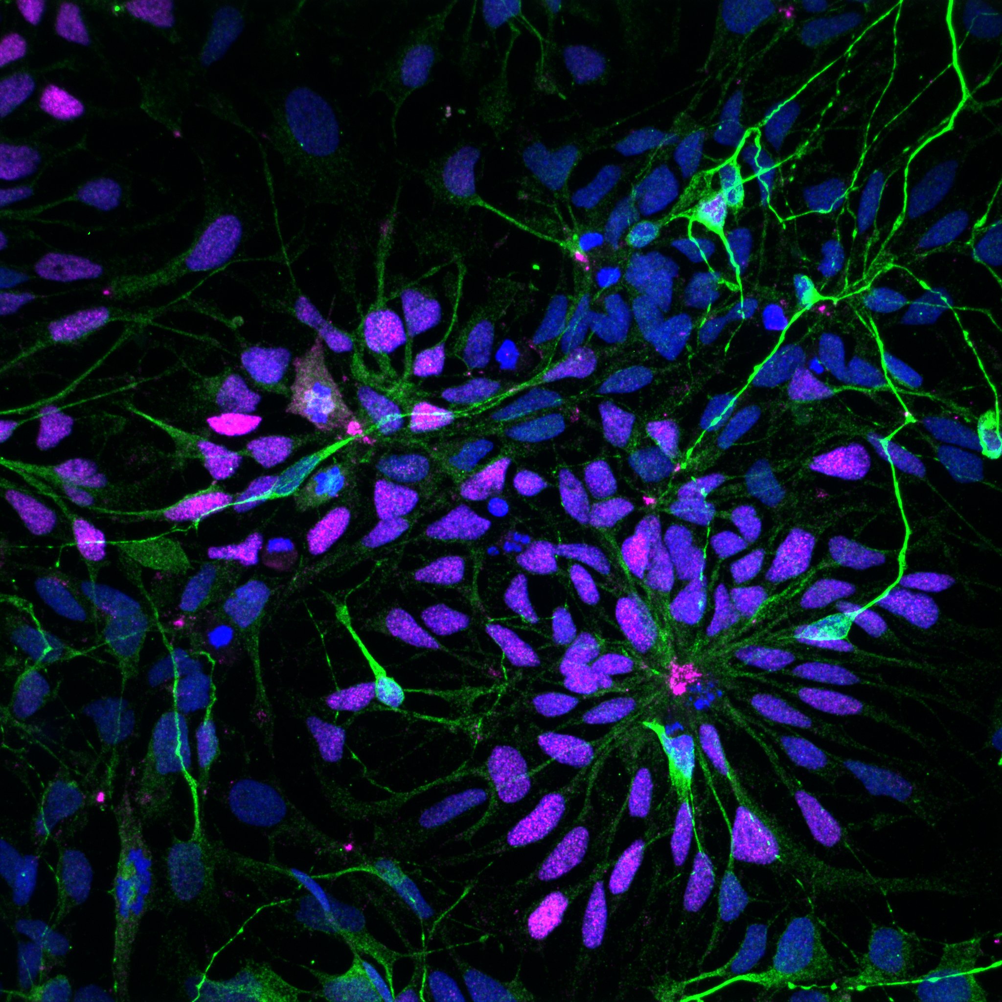
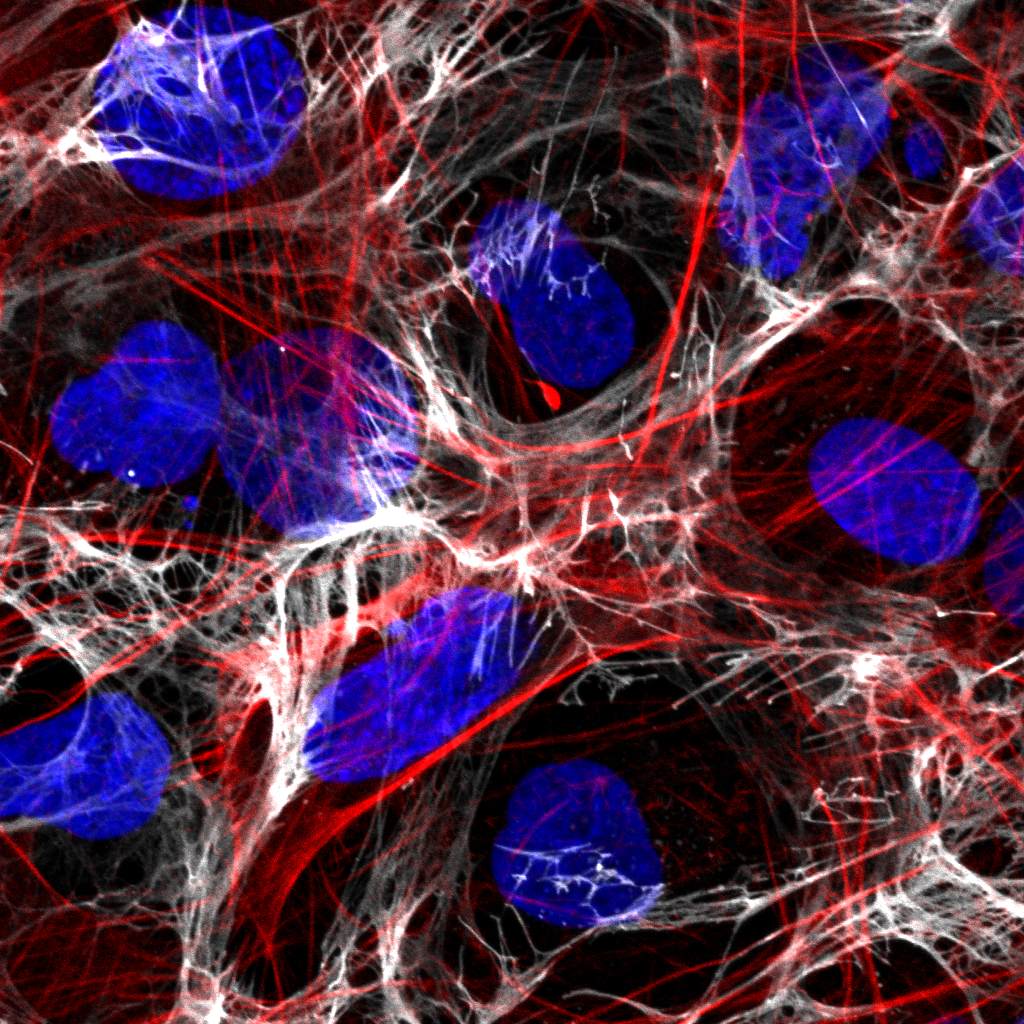
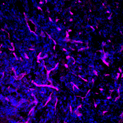
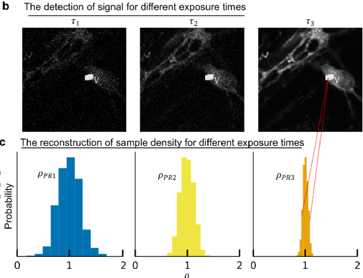
[Imaging] - Banner

Imaging

[Imaging] - Introduction

[Imaging] - Equipment 1

[Imaging] - Equipment 2

Divider

[Imaging] -Activities title

[Imaging] - Activities

GNAO1-related disorder - Bio Neuromolecular group, iPS cells and organoids

Pathogenic variants of the GNAO1 gene, encoding the alpha subunit of an inhibitory heterotrimeric guanine nucleotide-binding protein, have been linked to a neurodevelopmental disorder characterized by developmental delay, hypotonia, epilepsy and hyperkinetic movements. In this project we use iPSCs to generate cortical neurons, brain organoids, gastruloids and neuruloids to study the molecular mechanisms underlying this disorder, to test potential drugs and to develop new therapeutic approaches.


Benedetti MC, D'andrea T, Colantoni A, Silachev D, de Turris V, Boussadia Z, Babenko VA, Volovikov EA, Belikova L, Bogomazova AN, Pepponi R, Whye D, Buttermore ED, Tartaglia GG, Lagarkova MA, Katanaev VL, Musayev I, Martinelli S, Fucile S, Rosa A. Cortical neurons obtained from patient-derived iPSCs with GNAO1 p.G203R variant show altered differentiation and functional properties. Heliyon 2024 

The role of biological noise in cancer cell phenotypic plasticity - Bio Modeling Group

The concepts of phenotypic heterogeneity and plasticity in cell populations have gained considerable attention lately for their role in conferring cancer cells' resistance to treatments. This variability can stem from non-genetic factors such as the stochastic distribution of molecules during cell division. To address this issue systematically, we developed both experimental and computational tools to track the proliferation of cell populations and directly quantify the partitioning of various cellular components. Our approach involves initially staining different cellular elements with fluorescent markers, sorting the stained cells, and subsequently monitoring the dynamics of the partitioning process via extensive fluorescence microscopy time-lapses

OSTEOMImetic 3D Microfluidic bioprinting of decellularised human bone and vascular allograft ink for skeletal regeneration - OSTEOMIMIC, Bio Printing Group

To support bone regeneration, metal implants and prothesis are ideal, but fail to offer physiological repair providing an evident mismatch in mechanical properties with the bone tissue. Decellularized tissue derived hydrogels have attracted significant interest due to their similarity in composition to native tissue. In our work we want to harness hydrogels derived from native human placenta and bone matrix to be used in combination with human skeletal stem cells and endothelial cells to ultimately promote and guided vascular bone regeneration

The neurological consequences of peripheral inflammation: delving into synaptic transmission and glial responses - Sapienza University

Inflammatory Bowel Diseases (IBD) affect gastrointestinal tracts with extra-intestinal effects, such as anxiety and depression. In particular, some brain areas, i.e. hippocampus, respond to bowel inflammation modulating neuronal activity and glia density. In fact, at the peak of bowel inflammation microglia and glutamatergic synaptic transmission are altered. It has been observed that astrocytes acquire a neurotoxic phenotype and blood-brain barrier (BBB) is more permeable with subsequent immune cells infiltration. The hypothesis is that bowel inflammation induces neuroinflammation and synaptic alterations in hippocampus. The aim of the project is to highlight cellular and molecular mechanisms that underlie the alterations observed in the brain in mouse model of bowel inflammation. 


Scaringi G., Basilico B., Ragozzino D. Laboratorio di biofisica, dip. di Fisiologia e Farmacologia "V. Erspamer", Sapienza University

Targeting RNA-binding proteins in ALS - Bio Neuromolecular group, iPS cells and organoids

RNA-binding proteins (RBPs) play multiple roles in RNA metabolism and their mutation, delocalization and/or altered expression have been proposed to cause familial and sporadic amyotrophic latrla sclerosis (ALS). In this project, human iPSC-derived motor neurons, skeletal muscle cells and neuromuscular organoids are used as in vitro model systems to study the role of the RBPs FUS, HuD/ELAVL4 and TDP-43 in ALS.


Garone, M. G., Salerno, D. & Rosa, A. Digital color-coded molecular barcoding reveals dysregulation of common FUS and FMRP targets in soma and neurites of ALS mutant motoneurons. Cell Death Discov 9, 33 (2023)


Garone, M. G. et al. ALS-related FUS mutations alter axon growth in motoneurons and affect HuD/ELAVL4 and FMRP activity. Commun Biol 4, 1025 (2021).


Garone, M. G. et al. Proteomics analysis of FUS mutant human motoneurons reveals altered regulation of cytoskeleton and other ALS-linked proteins via 3'UTR binding. Sci Rep 10, 11827 (2020).


De Santis, R. et al. Mutant FUS and ELAVL4 (HuD) Aberrant Crosstalk in Amyotrophic Lateral Sclerosis. Cell Rep 27, 3818–3831.e5 (2019).


Garone, M. G. et al. Conversion of Human Induced Pluripotent Stem Cells (iPSCs) into Functional Spinal and Cranial Motor Neurons Using PiggyBac Vectors. J Vis Exp (2019). doi:10.3791/59321


De Santis, R. et al. Direct conversion of human pluripotent stem cells into cranial motor neurons using a piggyBac vector. Stem Cell Research 29, 189–196 (2018).


De Santis, R. et al. FUS Mutant Human Motoneurons Display Altered Transcriptome and microRNA Pathways with Implications for ALS Pathogenesis. Stem Cell Reports 9, 1450–1462 (2017).


Lenzi, J. et al. Differentiation of control and ALS mutant human iPSCs into functional skeletal muscle cells, a tool for the study of neuromuscolar diseases. Stem Cell Research 17, 140–147 (2016).


Lenzi, J. et al. ALS mutant FUS proteins are recruited into stress granules in induced Pluripotent Stem Cells (iPSCs) derived motoneurons. Dis Model Mech 8, 755–766 (2015).

Developing nanotechnological tool for cell-targeted delivery for diagnostic or therapeutic purposes - Bio Neuromolecular, Synaptic Organoid NeuroGlia lab

Human ferritins have been extensively studied to be used as nanocarriers for diverse applications and could represent a convenient alternative for targeted delivery of imaging agents and drugs for diagnostic or therapeutic purposes. Our studies highlight the potential of functionalized ferritin nanocages loaded with the fluorescent probe BT1 as a promising method for specifically identifying tau tangles in retinal tissue, aiding in the clinical diagnosis of tauopathies. Furthermore, the encapsulation of other substrates within ferritin nanocages opens up potential applications in the field of nanomedicine.


Soloperto A, Quaglio D, Baiocco P, Romeo I, Mori M, Ardini M, Presutti C, Sannino I, Ghirga S, Iazzetti A, Ippoliti R, Ruocco G, Botta B, Ghirga F, Di Angelantonio S, Boffi A. (2022). Rational design and synthesis of a novel BODIPY-based probe for selective imaging of tau tangles in human iPSC-derived cortical neurons. Sci Rep. 12(1):5257


Benni I., Trabuco M.C., Di Stasio E., Arcovito A., Boffi A., Malatesta F., Bonamore A., De Panfilis S., De Turris V., Baiocco P. (2018). Excimer based fluorescent pyrene-ferritin conjugate for protein oligomerization studies and imaging in living cells, RSC Advances, 8, 12815-12822.


De Turris V., Cardoso Trabuco M., Peruzzi G., Boffi A., Testi C., Vallone B., Celeste Montemiglio L., Georges A.D., Calisti L., Benni I., Bonamore A., Baiocco P. (2017). Humanized archaeal ferritin as a tool for cell targeted delivery, Nanoscale, 9, 647-655.

On-Chip Investigation: a potential novel therapeutic tool and its role in orchestrating ovarian cancer growth and immune activation - Bio Printing Group

To date, the controlled spatial arrangement of multiple cell types of ovarian tumour microenvironment, i.e. cancer and immune cells, in a 3D niche is still unexplored. A 3D ovarian cancer (OCa) model is realised harnessing a 3D microfluidic platform. A 3D human OCa tissue is cultured in a circular microfluidic chamber, in communication with a microfluidic channel through a porous membrane. The latter is perfused with immune cells to simulate and study the dynamics of cell extravasation from the blood vessel and infiltration to the tissue. The effect of a potential novel therapeutic tool is investigated, quantitatively assessing multiple processes involved in tumour growth and immunogenicity.

Bioprinting meniscus implants harnessing microfluidic biofabrication technology - BUBBLES, Bio printing Group

With the aid of a new microfluidic-assisted bioprinting technology, we aim to print a meniscus implant mimicking its vascularised structure. This is pursued by tagging the compartimentalised bioprinted construct with Vascular Endothelial Growth Factor (VEGF) and by printing a gradient three-dimensional architecture, promoting a microscopically controlled angiogenesis. This in turn support the bioprinting of a meniscal-like structure with a differential angiogenesis guided by the spatial confinement of VEGF compound and cellular components

A 3D bioprinted model for the study of Kabuki syndrome - Bio Printing Group & University of Trento

Kabuki syndrome is a rare neuro-developmental disorder caused by variants in genes of histone modification such as H3 lysine 4 methylase (KMT2D). This project aims to characterize the effects of KMT2D mutations on neuronal development, using an iPSC-derived 3D neural model obtained with microfluidic bioprinting.

Controlling the induction/reversal of Epithelial to Mesenchymal Transition (EMT) - Sapienza University
The gut-brain axis cross-talk in physiological and pathological condition - Sapienza University

In recent years, several studies described the close relationship between the composition of gut microbiota and brain functions, highlighting the importance of gut-derived metabolites in mediating neuronal and glial cells cross-talk in physiological and pathological conditions. In this project we aim to study how microbiota alteration may affect cerebral tumors growth and progression, and try to identify the specific metabolites involved in this modulation.


Rosito M., Maqbool J., Reccagni A., Giampaoli O., Sciubba F., Antonangeli F., Scavizzi F., Raspa M., Cordella F., Tondo L., Di Angelantonio S., Trettel F. , Miccheli A., D’Alessandro G., Limatola C.. Antibiotics treatment promotes vasculogenesis in the brain of glioma-bearing mice (2024). Cell death and disease - in press

Physics-informed deep neural network for image denoising - Bio Photonics group - Optical Design & Super-Resolution

Harnessing the power of artificial intelligence (AI), our research addresses challenges in microscopy. We focus on improving microscopic images through techniques such as reducing noise, enhancing contrast, and refining resolution. Our methods, including convolutional neural networks (CNNs) and residual neural networks (RNNs), cleverly boost signal-to-noise ratios. These networks, integral to machine learning, excel at extracting intricate details from complex biological samples. 


Xypakis E., de Turris V., Gala F., Ruocco G., Leonetti M. (2023). Physics-informed deep neural network for image denoising, Optics Express, 31, 43838-43849.

Pan-neuronal calcium imaging in C. elegans - C. elegans group

Volumetric calcium imaging recordings of C. elegans head neurons under different conditions for measuring the neuronal activity at single-cell resolution. We leverage the rapid acquisition rates of the spinning disk and a fast piezo stage to record entire volumes at a rate of 3 Hz. Nematodes are confined under the field of view through a microchip that enables the administration of chemical and mechanical stimuli. This setup enables capturing the neuronal activity of most head neurons, including those significantly contributing to the nervous system's collective dynamics.


Lanza E., Lucente V., Nicoletti M., Schwartz S., Cavallo I.F., Caprini D., Connor C.W., Saifuddin M.F.A., Miller J.M, L’Etoile N.D., Folli V. (2024) See Elegans: Simple-to-use, accurate, and automatic 3D detection of neural activity from densely packed neurons. PLOS ONE, 19(3), e0300628

Microfluidic 3D Printing of Emulsion Ink for Engineering Porous Functionally Graded Materials - Bio Printing Group

An innovative approach to 3D printing for the production of polymeric materials characterised by trabecular porosity and functionally graded architecture was developed for multi-interface tissue fabrication. These types of materials exhibit local variations in mechanical and chemical composition of the matrix in three-dimensional space. We engineered a library of functionally graded materials (FGMs) capable of driving cell-specific activities in 3D towards complex tissue development and modelling


M. Marcotulli,  M. C. Tirelli,  M. Volpi,  J. Jaroszewicz,  C. Scognamiglio,  P. Kasprzycki,  K. Karnowski,  W. Święszkowski,  G. Ruocco,  M. Costantini,  G. Cidonio,  A. Barbetta. (2023)  Microfluidic 3D Printing of Emulsion Ink for Engineering Porous Functionally Graded Materials. Adv. Mater. Technol.,  8, 2201244.

Integrative approach for modelling neurodegeneration using patient-derived iPSCs - Bio Neuromolecular group, Neurodegeneration and protein aggregation

In our effort of unraveling the complexities of neurodegenerative diseases, our primary objective is the establishment of robust and reliable in vitro models. The use of iPSCs allows us to replicate the unique genetic profiles of individual patients, providing a personalized dimension to our investigations. Furthermore, the incorporation of organoid cultures adds a three-dimensional aspect, better mimicking the intricate cellular architecture of the human brain. An additional component of our methodology involves microfluidics, which enables precise control over the microenvironment, facilitating the recreation of physiological conditions within our in vitro models. By combining these advanced techniques, we aim to bridge the gap between traditional in vitro models and the complex in vivo environment, paving the way for a deeper understanding of the molecular events contributing to neurodegenerative processes.

Protein aggregation and protein-protein interaction as therapeutic targets for neurodegenerative diseases - Bio Neuromolecular group, Neurodegeneration and protein aggregation

Many neurodegenerative diseases stem from compromised proteostasis and mutations leading to protein aggregation. Our primary objective is to elucidate the mechanisms underlying aggregation, aiming to understand potential interactions between the aggregates and other proteins that give rise to toxic functions. To achieve this, we utilize imaging techniques on our in vitro disease models, focusing specifically on Amyotrophic Lateral Sclerosis and Huntington's Disease. Through this comprehensive approach, we aim to develop effective peptides that can act as therapeutics to disrupt pathogenic interactions. Furthermore, we are investigating the potential of drug repurposing as a strategy to alleviate altered proteostasis and mitigate protein aggregation.

Bioengineered hydrogels as 3D Cultured Model - CNR Nanotec

Bioengineered hydrogels represent physiologically relevant platforms for cell behavior studies in the tissue engineering and regenerative medicine fields, as well as in in vitro disease models. Hyaluronic acid (HA) is an ideal platform since it is a natural biocompatible polymer that is widely used to study cellular crosstalk, cell adhesion and cell proliferation, and is one of the major components of the extracellular matrix (ECM). The morphological evolution processes and proliferation rates of glioblastoma cells (U251-MG) cultured on HA-MA surfaces were evaluated by comparing 2D structures with 3D structures. Moreover cell viability assays, evaluation of mitochondrial metabolism and morphological studies has been performed.


Ursini O., Grieco M., Sappino C., Capodilupo A.L., Giannitelli S.M., Mauri E., Bucciarelli A., Coricciati C., de Turris V., Gigli G., Moroni L., Cortese B. (2023). Modulation of Methacrylated Hyaluronic Acid Hydrogels Enables Their Use as 3D Cultured Model, Gels, 9.

Collective behavior and self-organization - Bio Modeling Group

Systems biology is an interdisciplinary field that aims to understand biological systems as a whole, rather than just studying individual components. It uses computational algorithms and modeling techniques to analyze complex cellular processes and interactions, helping researchers gain a deeper understanding of how these systems work and how they can be manipulated to improve human health.


Miotto M., Rosito M., Paoluzzi M., de Turris V., Folli V., Leonetti M., Ruocco G., Rosa A., Gosti G. (2023). Collective behavior and self-organization in neural rosette morphogenesis, Frontiers in Cell and Developmental Biology, 11.

3D structure of the human centromere in mitotic chromosomes – Sapienza University

The human centromere comprises large arrays of repetitive α-satellite DNA at the primary constriction of mitotic chromosomes. In addition, centromeres are epigenetically specified by the centromere-specific histone H3 variant CENP-A that supports kinetochore assembly to enable chromosome segregation. Because CENP-A is bound to only a fraction of the α-satellite elements within the megabase-sized centromere DNA, correlating the three-dimensional (3D) organization of α-satellite DNA and CENP-A remains elusive.


Di Tommaso E., de Turris V., Choppakatla P., Funabiki H., Giunta S. (2023). Visualization of the three-dimensional structure of the human centromere in mitotic chromosomes by superresolution microscopy, Molecular Biology of the Cell, 34.

An in vivo humanized model to study homing and sequestration of Plasmodium falciparum transmission stages in the bone marrow – Sapienza University & Istituto Superiore di Sanità

They generated a novel experimental system based on the infusion of immature P. falciparum gametocytes into immunocompromised mice carrying chimeric ectopic ossicles whose stromal and bone compartments derive from human osteoprogenitor cells. This model represents a powerful tool to study BM function and the interplay essential for parasite transmission in P. falciparum malaria and can be extended to study other infections in which the human BM plays a role.


Donsante S., Siciliano G., Ciardo M., Palmisano B., Messina V., de Turris V., Farinacci G., Serafini M., Silvestrini F., Corsi A., Riminucci M., Alano P. (2023). An in vivo humanized model to study homing and sequestration of Plasmodium falciparum transmission stages in the bone marrow, Frontiers in cellular and infection microbiology, 13.

Microglia and neuronal spine morphology - Sapienza University

Microglia cells are active players in regulating synaptic development and plasticity in the brain. Microglia contribute to normal synaptic functioning in the adult brain but how they influence the normal functioning of synapses is largely unknown. We support various projects by acquiring and post-processing by deconvolution (Huygens software) mouse brain confocal images from different treatments and condition. These allow for the analysis of microglia and dendritic spine density and morphology in different contest such as microglia pharmaceutical depletion, interactions with immune cells in physiological and pathological conditions such sleep and brain tumors.


Garofalo S., dip. di Fisiologia e Farmacologia "V. Erspamer", Sapienza University


Basilico B., Ferrucci L., Ratano P., Golia M.T., Grimaldi A., Rosito M., Ferretti V., Reverte I., Sanchini C., Marrone M.C., Giubettini M., De Turris V., Salerno D., Garofalo S., St-Pierre M.-K., Carrier M., Renzi M., Pagani F., Modi B., Raspa M., Scavizzi F., Gross C.T., Marinelli S., Tremblay M.-E., Caprioli D., Maggi L., Limatola C., Di Angelantonio S., Ragozzino D. (2022). Microglia control glutamatergic synapses in the adult mouse hippocampus, GLIA, 70, 173-195.

Imaging a vessel on-a-chip for drug delivery studies

A local uptake of drugs at the target requires drugs diffusion across the endothelial barrier lining the blood vessel walls and through the surrounding tissue. A vessel-on-a-chip is exploited to investigate and quantify the effect of ultrasound-excited microbubbles -a process named cavitation- on endothelial integrity. Microbubbles amplify the ultrasound effect, leading to the formation of inter-endothelial gaps that cause barrier permeabilization and enhance drug delivery. The proposed integrated platform allows for precise and repeatable in vitro measurements of cavitation-enhanced endothelium permeability and shows potential for validating irradiation protocols for in vivo applications


A Microfluidic Platform for Cavitation-Enhanced Drug Delivery, Grisanti G., Caprini D., Sinibaldi G., Scognamiglio C. , Silvani G. ,Peruzzi G., and Casciola C.M., Micromachines 2021


Reversible Cavitation-Induced Junctional Opening in an Artificial Endothelial Layer, Silvani G., Scognamiglio C., Caprini D., Marino L., Chinappi M., Sinibaldi G., Peruzzi G., Kiani F.  M., and Casciola C. M., Small 2019

Lipid Rafts and Infection - Istituto Superiore di Sanità

Lipid rafts are small, highly dynamic and tightly ordered plasma membrane microdomains, enriched in cholesterol, glycosphingolipids, glycosylphosphatidylinositol (GPI)-linked proteins and signaling-related molecules, that play a major role in regulation of protein sorting and organization within cell membranes. In particular, lipid rafts can establish specialized membrane clusters where diverse cellular receptors are co-localized, concentrated and segregated with partners of their downstream signaling pathways and are crucially involved in coordination of cell signal transduction. In recent years, raft microdomains have emerged as crucial surface platforms through which several bacterial, protozoan and viral pathogens can interact with host phagocytes to trigger or modulate the early anti-infectious innate immune response and the ensuing adaptive immune response.


Peruzzu D., Amendola A., Venturi G., de Turris V., Marsili G., Fortuna C., Fecchi K., Gagliardi M.C. (2022). Zika Virus Exploits Lipid Rafts to Infect Host Cells, Viruses, 14.


Mariotti S., Teloni R., de Turris V., Pardini M., Peruzzu D., Fecchi K., Nisini R., Gagliardi M.C. (2020). Amphotericin B inhibits mycobacterium tuberculosis infection of human alveolar type II epithelial A549 cells, Antimicrobial Agents and Chemotherapy, 64.


De Turris V., Teloni R., Chiani P., Bromuro C., Mariotti S., Pardini M., Nisini R., Torosantucci A., Gagliardi M.C. (2015). Candida albicans targets a lipid raft/dectin-1 platform to enter human monocytes and induce antigen specific T cell responses, PLoS ONE, 10.

Development of reliable models of pediatric low-grade gliomas - Sapienza University

Pediatric low-grade gliomas (pLGGs), the most frequent pediatric brain tumors, include different entities harboring distinct histological and molecular features. A major limitation in the development of treatments for these tumors is the absence of reliable in vitro models that would allow a better understanding of the molecular mechanisms that support their growth. Surgical excision is the primary treatment method and the extent of resection represents one of the strongest prognostic factors. pLGGs that cannot be completely resected are prone to recur and associated with relapses and extensive morbidities, thus remaining a major clinical challenge. The development of reliable 2D and 3D models of pLGGs is of utmost importance to deepen the molecular knowledge about this tumors with the aim to develop new and more effective therapeutical strategies.


Chiacchiarini M., Besharat Z.M., Carai A., Miele E., Del Baldo G., Mastronuzzi A., Catanzaro G.*,§, Ferretti E. *,§ Pediatric low-grade gliomas: molecular characterization of patient-derived cellular models. Child’s Nervous System, 2021 Mar; 37(3):771-778